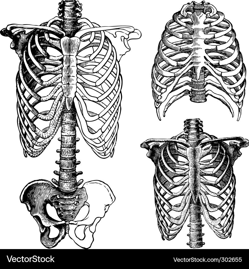
Anatomy Of Chest Bones - The scapula, or shoulder blade, is an approximately triangular shaped bone.
byAdmin•
0
Anatomy Of Chest Bones - The scapula, or shoulder blade, is an approximately triangular shaped bone.. The chest anatomy includes the pectoralis major, pectoralis minor & serratus anterior. Language and terminology for the study of the anatomy of the thorax. The medial anterior chest is defined by the sternum, which consists of 3 flat polygonal bones: The wrist consists of multiple joints where the bones of the arm and hand meet. The ribs meet at an acute angle at the sternum, the costal cartilages thicken like beads at points of their transition to bones (rachitic beads).
Long bones are mostly located in the appendicular skeleton and include bones in the lower limbs (the tibia, fibula, femur, metatarsals, and phalanges) and bones in the upper limbs (the humerus, radius, ulna, metacarpals. A collection of anatomy notes covering the key anatomy concepts that medical students need to learn. The medial anterior chest is defined by the sternum, which consists of 3 flat polygonal bones: All of the anatomical and important histological facts about the bones, together with the clinical relations, are going to be desrcibed in this article. There also are bands of fibrous connective tissue—the ligaments and the tendons—in intimate relationship with the parts of the skeleton.
Anatomical Chest Drawings Royalty Free Vector Image from cdn1.vectorstock.com Bones are mostly made of the protein collagen , which forms a soft framework. This webpage presents the anatomical structures found on wrist mri. Human chest bone structure parts of the chest bones. A collection of anatomy notes covering the key anatomy concepts that medical students need to learn. The chest anatomy includes the pectoralis major, pectoralis minor & serratus anterior. Long bones are categorised by their tubular shaft (diaphysis) with a rounded end (epiphysis) on each end. Anatomy of the chest wall. Sesamoid bones are generally small, flat and have an apex at one end.
This framework consists of many individual bones and cartilages.
The medial anterior chest is defined by the sternum, which consists of 3 flat polygonal bones: Sesamoid bones are generally small, flat and have an apex at one end. They are always longer than they are wide the vertebrae are irregular bones. We hope you will use this picture in the study and helping chest and abdominal cavities with some organs removed. These bones form by the thickening of a. What can you label/identify on the nmt exam. There also are bands of fibrous connective tissue—the ligaments and the tendons—in intimate relationship with the parts of the skeleton. The manubrium, sternal body, and xiphoid process. Compare the nuclear medicine scans to anatomical diagrams. It describes the theatre of events. Despite this it is easy to overlook important abnormalities of the bones which may be very subtle. Your rib cage, for example, acts like a shield around your chest to protect important organs inside such as your lungs and heart. It originates at your clavicle, ribs, and sternum, and inserts into the upper portion of your humerus (upper arm bone from elbow to shoulder.)
Anatomy of the chest wall. Atlas of wrist mri anatomy. Despite this it is easy to overlook important abnormalities of the bones which may be very subtle. Your rib cage, for example, acts like a shield around your chest to protect important organs inside such as your lungs and heart. Atlas of anatomy of the human body:
Rib Cage Anatomy Labeled Vector Illustration Diagram Medical Human Chest Skeletal Bone Structure Model Numbered Ribs Sternum Cartilage Parts And Clavicular Articulation Health Care Education Royalty Free Cliparts Vectors And Stock Illustration from previews.123rf.com Your rib cage, for example, acts like a shield around your chest to protect important organs inside such as your lungs and heart. There also are bands of fibrous connective tissue—the ligaments and the tendons—in intimate relationship with the parts of the skeleton. It, essentially, floats off of the back of the chest, as it is connected to the body primarily by muscle. What can you label/identify on the nmt exam. They are always longer than they are wide the vertebrae are irregular bones. A collection of anatomy notes covering the key anatomy concepts that medical students need to learn. Atlas of wrist mri anatomy. We hope you will use this picture in the study and helping chest and abdominal cavities with some organs removed.
Bone basics and bone anatomy.
Ground substance and collagen fibers create a matrix that contains. Breast bone anatomy human breast bone anatomy bone anatomy sternum | chest bone : There also are bands of fibrous connective tissue—the ligaments and the tendons—in intimate relationship with the parts of the skeleton. Have you ever seen fossil remains of dinosaur and ancient human bones in textbooks, television, or in person at a museum? Identify the following structures on the lateral chest radiograph: A collection of anatomy notes covering the key anatomy concepts that medical students need to learn. It can help you understand our world more detailed and specific. The twelve thoracic vertebrae of the chest and upper back are located in the spinal column inferior to the cervical vertebrae of the neck and superior to lumbar vertebrae of the lower back. It is comprised of many bones, formed by intramembranous ossification, which are joined together by sutures (fibrous joints). These bones form by the thickening of a. The scapula, or shoulder blade, is an approximately triangular shaped bone. Long bones are categorised by their tubular shaft (diaphysis) with a rounded end (epiphysis) on each end. Bone structure on plain studio background.human anatomy chest from low angle.
It describes the theatre of events. Compare the nuclear medicine scans to anatomical diagrams. Spot views were taken of the chest, spine, hand, and foot. Atlas of anatomy of the human body: Parts of the chest bones for many, the chest is made up of a.
Shoulder And Chest Anatomy Artwork Stock Image C020 0121 Science Photo Library from media.sciencephoto.com The two bones are joined at a slight angle that protrudes anteriorly (sternal angle, angle of louis). Language and terminology for the study of the anatomy of the thorax. Bones are mostly made of the protein collagen , which forms a soft framework. The ribs meet at an acute angle at the sternum, the costal cartilages thicken like beads at points of their transition to bones (rachitic beads). The largest bone in the human body is the thighbone or femur, and the smallest is the stapes in the middle ear, which are just 3 millimeters (mm) long. A bone is a somatic structure that is comprised of calcified connective tissue. It is comprised of many bones, formed by intramembranous ossification, which are joined together by sutures (fibrous joints). Despite this it is easy to overlook important abnormalities of the bones which may be very subtle.
Anatomy is the amazing science.
The largest bone in the human body is the thighbone or femur, and the smallest is the stapes in the middle ear, which are just 3 millimeters (mm) long. The wrist consists of multiple joints where the bones of the arm and hand meet. O bones—spine, ribs, clavicles, scapulae, humeri. A collection of anatomy notes covering the key anatomy concepts that medical students need to learn. We hope you will use this picture in the study and helping chest and abdominal cavities with some organs removed. What can you label/identify on the nmt exam. Anatomy of the chest wall. Bones of the chest and upper back (posterior view). Long bones function to support the weight of the body and facilitate movement. Ground substance and collagen fibers create a matrix that contains. Anatomy is the amazing science. There also are bands of fibrous connective tissue—the ligaments and the tendons—in intimate relationship with the parts of the skeleton. When a patient flexes the neck forward, the prominent process is usually that of the 7th cervical.
They are always longer than they are wide the vertebrae are irregular bones anatomy of chest. These joints fuse together in adulthood, thus permitting brain growth during adolescence.